A novel radiotracer paves the way for effective live imaging of α-synuclein deposits in patients with Parkinson’s and related disease.
Many neurodegenerative disorders, including Parkinson’s disease (PD) and dementia with Lewy bodies (DLB), are very difficult to diagnose and study in living subjects. Now, researchers from Japan have successfully developed a chemical radiotracer that binds to α-synuclein fibrils, harmful protein aggregates typically seen in PD and DLB pathologies. Their efforts could make these diseases diagnosable based on background pathology via tomography and help with the development of effective treatments and drugs.
In countries with an aging population, neurodegenerative disorders such as Parkinson’s disease (PD) and dementia with Lewy bodies (DLB) are becoming more prevalent. These conditions, for which no definitive cure currently exists, are extremely difficult to diagnose accurately while the affected person is still alive. This has proved to be a major roadblock for researchers seeking to assess potential therapies, since they have no conclusive way to check whether their proposed treatment is truly working in either patients or animal models.
Like Alzheimer’s disease, a hallmark symptom in PD and DLB is the abnormal accumulation of certain protein structures. In PD and DLB, these aggregates are known as α-synuclein fibrils, which spread throughout the brain and cause damage to neural pathways. Many scientists are trying to develop chemical agents that can selectively bind to α-synuclein and make it detectable via techniques such as positron emission tomography (PET). While a few studies have seen some success with this approach, there are still no PET tracers for α-synuclein that can be used in clinical practice to diagnose PD and DLB in living patients. This is mostly because α-synuclein deposits are small and not overly abundant.
Recently, as reported in a new study published in the journal Neuron on June 05, 2024, a team of scientists from Japan has designed a potentially game-changing PET tracer for α-synuclein. The team was led by Senior Researcher Hironobu Endo from the Advanced Neuroimaging Center, Institute for Quantum Medical Science, and included Dr. Maiko Ono from the Institute for Quantum Life Science and Dr. Makoto Higuchi from the Advanced Neuroimaging Center, Institute for Quantum Medical Science, all based at the National Institutes for Quantum Science and Technology (QST).
The researchers first screened various potentially useful compounds that were selected based on prior knowledge. After a series of preliminary experiments in vitro, they found that a compound derived from PBB3—typically used to detect tau protein aggregates in Alzheimer’s disease—was quite useful in visualizing α-synuclein. The team labeled this chemical, (E)-1-fluoro-3-((2-(4-(6-(methylamino)pyridine-3-yl)but-1-en-3-yn-1-yl)benzo[d]thiazol-6-yl)oxy)propan-2-ol, as C05-05.
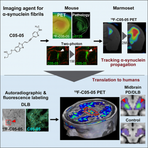
Through extensive tests performed in two different animal models, namely mice and marmosets, the researchers showcased the high binding affinity and selectivity of C05-05 to α-synuclein. More specifically, C05-05 was used to analyze the quantity and distribution of these harmful protein deposits in various areas of the brain at different stages of disease progression. Remarkably, C05-05 could also be used at the single-cell level via two-photon microscopy, making it a powerful tool for understanding the fine details of how α-synuclein aggregates damage neurons. “Our tracer enabled multi-scale imaging of α-synuclein depositions in living animal models, which is crucial for translational research and development,” remarks Dr. Higuchi.
Next, the researchers tested the clinical potential of C05-05 as a PET tracer in humans. To this end, they recruited 10 patients with symptoms of PD and DLB, as well as eight healthy people as controls. In line with the experimental results on animal models, the researchers observed that the proposed compound was useful for visualizing α-synuclein depositions, especially in a deep structure known as the midbrain. The intensity of C05-05 signals in this region was enhanced in patients with PD and DLB compared to that of the control group, showcasing the first demonstration of α-synuclein imaging in live humans.
These findings have important implications for both drug development and diagnostics, as Dr. Endo highlights: “In vivo imaging techniques for detecting α-synuclein aggregates could provide definitive information on the disease diagnosis based on pathology and would be very useful to evaluate the efficacy of candidate drugs targeting α-synuclein pathologies at non-clinical and subsequently clinical levels.”
While there is still much work to do before we reach effective therapies for PD and related disorders, more engineering breakthroughs are underway. “QST is committed to the development of innovative head-dedicated PET scanners suitable for high-resolution, high-sensitivity detection of α-synuclein deposits in small brain structures like the midbrain. Hopefully, these technologies will open doors to a diagnostic package approved by regulatory agencies within five years,” speculates Dr. Higuchi.
Let us all hope for a brighter future where PD and other neurodegenerative disorders can be diagnosed and stopped in their tracks early on.

Image title: Multi-scale imaging of harmful protein aggregates in live patients with PD and DLB.
Image caption: Researchers from Japan developed a novel tracer compound that binds to α-synuclein aggregates, making them easy to detect in PET scans and two-photon microscopy. This chemical agent could greatly facilitate the diagnosis of complex neurodegenerative diseases, such as PD and DLB, in living patients and help during the testing and development phase of potential drug candidates.
Image credit: Hironobu Endo from the National Institutes for QST (https://www.cell.com/neuron/fulltext/S0896-6273(24)00332-5)
License type: CC BY-NC 4.0
Usage restrictions: Credit must be given to the creator. Only non-commercial uses of the work are permitted.
Reference
|
Imaging α-synuclein pathologies in animal models and patients with Parkinson’s and related diseases |
|
|
Journal |
Neuron |
|
DOI: |
About National Institutes for Quantum Science and Technology, Japan
The National Institutes for Quantum Science and Technology (QST) was established in April 2016 to promote quantum science and technology in a comprehensive and integrated manner. The new organization was formed from the merger of the National Institute of Radiological Sciences (NIRS) with certain operations that were previously undertaken by the Japan Atomic Energy Agency (JAEA).
QST is committed to advancing quantum science and technology, creating world-leading research and development platforms, and exploring new fields, thereby achieving significant academic, social, and economic impacts.
Website: https://www.qst.go.jp/site/qst-english/
About Senior Researcher Hironobu Endo from National Institutes for Quantum Science and Technology (QST), Japan
Hironobu Endo is a Senior Researcher at the Brain Disorder Translation Research Group at the Advanced Neuroimaging Center, Institute for Quantum Medical Science, National Institutes for Quantum Science and Technology (QST) in Japan. He specializes in neurological disorders and cutting-edge brain and neural imaging techniques, including functional brain imaging. He has published over 50 research papers on these topics, which have been cited over 300 times.
Funding information
This study was supported in part by MEXT KAKENHI grant number JP22K07529; by AMED under grant numbers JP18dm0207018, JP21zf0127004, JP21wm0425015, JP22dm0207072,
JP21dk027046, and JP22dk0207063; by JST moonshot R&D grant number JPMJMS2024, and by the Collaborative Research Project (2021-201907) of the Brain Research Institute, Niigata University.
Media contact:
Public Relations Section
Department of Management and Planning, QST
Tel: +81-43-206-3026 Email: info@qst.go.jp
