
Department of Accelerator and Medical Physics are operating and improving the synchrotron accelerators for heavy-ion cancer therapy, the cyclotron accelerators for production of radioisotopes as used for the diagnosis and treatment of cancer and dementia, and the electrostatic accelerator for PIXE elemental analysis and a microbeam cell irradiation. We are also conducting research and development of medical physics, radiation biology, and accelerator physics using these accelerators, and the outcome of our research activities contributes to improve people's lives in fields of healthcare.
organization
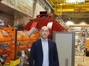
Iwata Yoshiyuki Director
Research Group
- Advanced Particle Therapy System Research Group(Mizushima Kota)
- Therapeutic Beam Research Group(Inaniwa Taku)
- Treatment System Research Group(Mori Shinichiro)
- Radiation Effect Research Group(Shimokawa Takashi)
- Beam Delivery System Research Group(Yonai Shunsuke)
- HIMAC Operation Section(Katagiri Ken)
- Cyclotron Operation Section(Wakui Takashi)
- Electrostatic Accelerator Operation Section(Oikawa Masakazu)