
VISUALIZATION
Ion beam can visualize phenomena in a living thing as it is.
Elemental mapping in a microscopic area
Particle induced x-ray emission spectrometry (PIXE) is a popular analytical method for various applications, since it is nondestructive, multi-elemental, and highly sensitive for the determination of trace elements. Its combination with an external scanning ion microbeam provides microscopic elemental analysis in atmospheric environment. This novel technique called Micro-PIXE is developed at TIARA and used to visualize elemental distributions in biological cells. The study is expected to reveal uptake mechanisms of medicine in cellular level.
How is it visuallzed?
An ion beam can be focused in a spot as small as 1mm in a specimen with a set of quadrupole magnets.
Characteristic X-rays are emitted from the atoms that collided with incident beam ions.
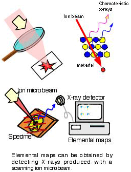

What is expected?
Micro-PIXE technique is a powerful tool not only for bio-medical research, but also for dentistry, environmental science and geology, because of its high sensitivity and good spatial resolution. In dentistry, uptake and transfer of fluorine from dental materials to teeth are measured. For environmental study, elemental concentrations of individual aerosol particles can be known. Trace-element distributions in geological samples are also investigated.
Movement of molecules and ions in a plant
Micro-PIXE technique is a powerful tool not only for bio-medical research, but also for dentistry, environmental science and geology, because of its high sensitivity and good spatial resolution. In dentistry, uptake and transfer of fluorine from dental materials to teeth are measured. For environmental study, elemental concentrations of individual aerosol particles can be known. Trace-element distributions in geological samples are also investigated.
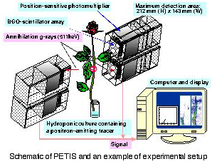
Positron-emitting radioisotopes are used for this purpose. For instance, the radioisotope 11C, which is produced by bombarding a nitrogen-gas target with a proton beam, decays by positron emission followed by positron-electron annihilation with the emission of two γ-rays at 180°. The γ-rays have sufficient energy (511 keV) to penetrate several cm of plant tissue or several ten meters of air, and can be detected in time coincidence by two detectors. This makes it possible to localize the source in a two-dimensional sample without touching the sample. Based on this principle, the Positron Emitting Tracer Imaging System (PETIS) has been developed.
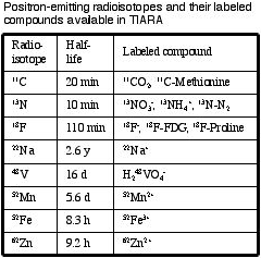
PETIS is the world's first system that is designed for real- time imaging of the distribution of a positron-emitting radioisotope in a plant. From consecutive image data stored in its computer memory, the movement of molecules and ions can be studied. Meanwhile, plant research needs various compounds labeled with positron-emitting radioisotopes. Most positron-emitting radioisotopes, however, are so short-lived, except for 22Na, that they have to be produced at the site for experiments using PETIS. The TIARA facility provides these conditions, including labeled compounds of nutrients and amino acids, and PETISs installed in the controlled environment chambers equipped with radioactive-gas-handling systems.
What has been and will be performed with this new technique?
Uptake and translocation of nutrients, fed from roots or leaves, and products in assimilation have been visualized for higher plants such as rice, barley and bean. For example, the visualization of the translocation of 11C-methionine has revealed an important part of the mechanism for a barley to absorb iron from alkali soil; this amino acid works as a precursor of the chelating agent excreted from the roots to absorb iron insolubilized in alkali soil. With PETIS, you can study behavior of compounds in a living plant under different environmental conditions. Effects of elevated CO2 concentrations on crop harvesting, possibilities of removal of toxic substances by plants, and others related to the security of food resources or the conservation of the environment will be the subjects of research.
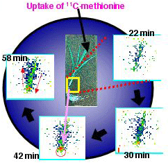
The world’s first real-time image of the translocation of an amino acid in a higher plant: 11C-methionine moved from a leaf of barley, the cut tip of which was dipped into a solution containing 11C-methionine, to another leaf.