
Updated:13 Feb. 2015
Team Leader:
Yumiko SUTO
Members:
- Yoshio TAKASHIMA
- Miho AKIYAMA
- Makiko OWAKI
- Momoki HIRAI
Inter-Institutional Collaborators:
- Naoki OSADA
(National Institute of Genetics, Mishima) - Ryuichi SAKATE
(National Institute of Biomedical Innovation, Osaka) - Shuhei MANO
(The Institute of Statistical Mathematics, Tokyo)
New Publication
Assessing the Applicability of FISH-based Prematurely Condensed Dicentric Chromosome Assay in Triage Biodosimetry.Health Physics,2015 March,108(3): 371-376.
- Assessing the applicability of FISH-based prematurely condensed dicentric chromosome assay in triage biodosimetry.
- Supplementary Technical Note[PDFファイル/30KB]
Radiation Cytogenetics Gallery:
Chromosome Aberration Data:Table 1
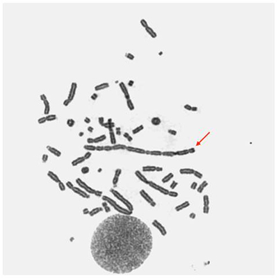
Fig. 1 A "deca"centric chromosome (arrow) in a human peripheral blood lymphocyte induced by 15-Gy gamma-irradiation.
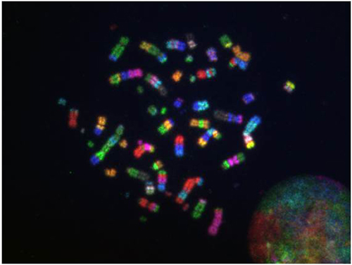
Fig. 2 Complicated chromosomal rearrangements in a 20-Gy irradiated human peripheral blood lymphocyte (M-FISH).
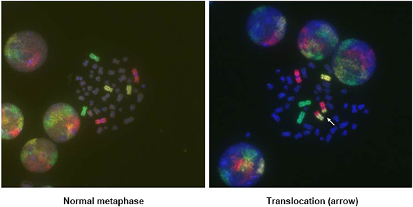
Fig. 3-1 Chromosomal rearrangements detected by 3-color FISH.
(#1, red; #2, green; #4, yellow)
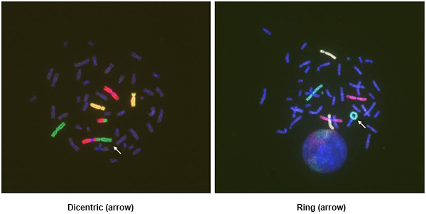
Fig. 3-2 Chromosomal rearrangements detected by 3-color FISH.
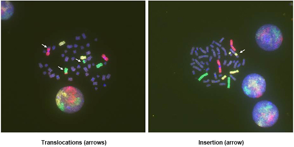
Fig. 3-3 Chromosomal rearrangements detected by 3-color FISH.
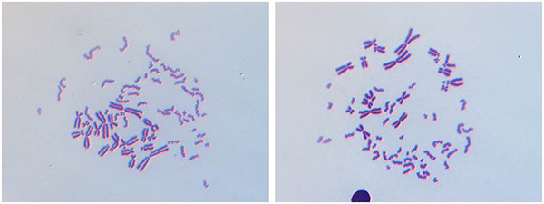
Fig. 4 Cell fusion-mediated premature chromosome condensation (PCC) in human peripheral blood lymphocytes observed by differential interference contrast microscopy.
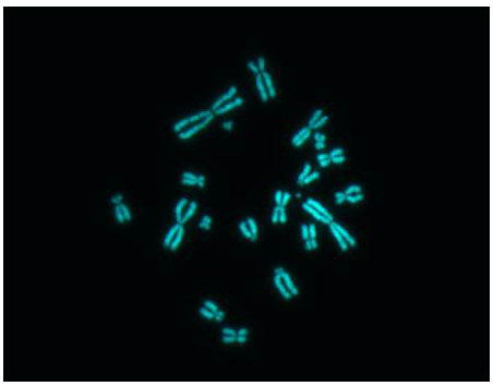
Fig. 5 Chinese Hamster Ovary metaphase cell (CHO-K1).
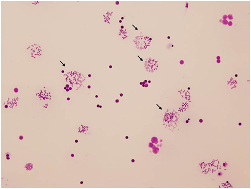
Fig. 6 Hybrid cells with prematurely condensed human chromosomes by conventional Giemsa-staining (arrows) (✕ 200 magnification).
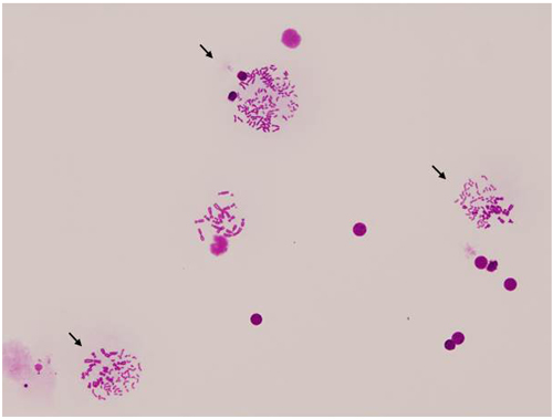
Fig. 7 Hybrid cells with prematurely condensed human chromosomes by conventional Giemsa-staining (arrows) (✕ 400 magnification)
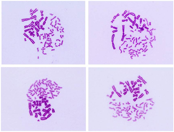
Fig. 8-1 Examples of PCC images (non-irradiated human lymphocytes).
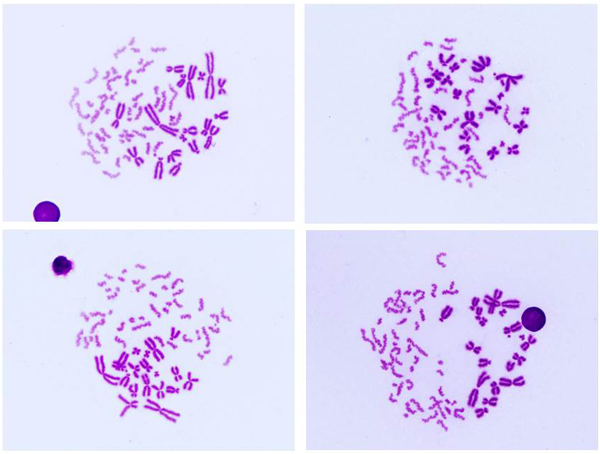
Fig. 8-2 Examples of PCC images (non-irradiated human lymphocytes).
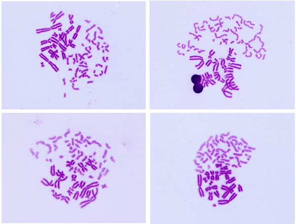
Fig. 8-3 Examples of PCC images (non-irradiated human lymphocytes).
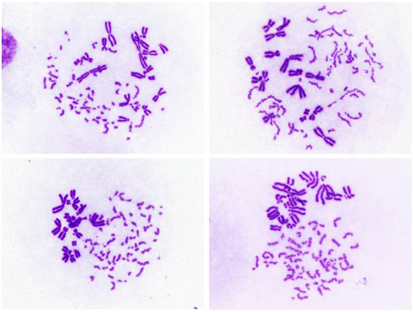
Fig. 9-1 Examples of PCC images (human lymphocytes hybridized with mitotic CHO cells immediately after 4-Gy gamma-irradiation).
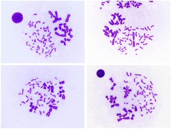
Fig. 9-2 Examples of PCC images (human lymphocytes hybridized with mitotic CHO cells immediately after 4-Gy gamma-irradiation).
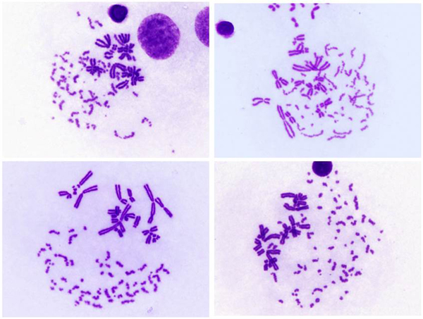
Fig. 9-3 Examples of PCC images (human lymphocytes hybridized with mitotic CHO cells immediately after 4-Gy gamma-irradiation).
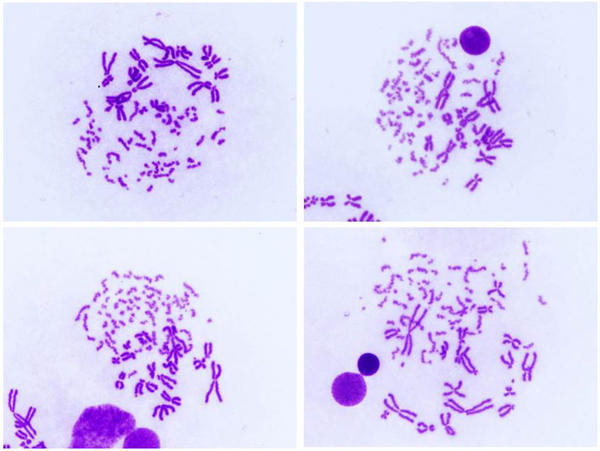
Fig. 9-4 Examples of PCC images (human lymphocytes hybridized with mitotic CHO cells immediately after 4-Gy gamma-irradiation).
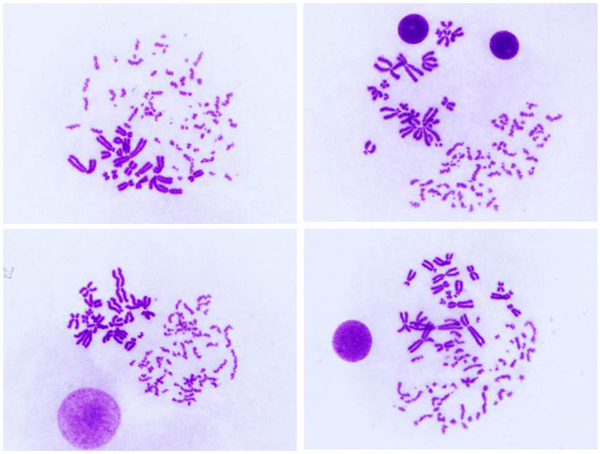
Fig. 9-5 Examples of PCC images (human lymphocytes hybridized with mitotic CHO cells immediately after 4-Gy gamma-irradiation).
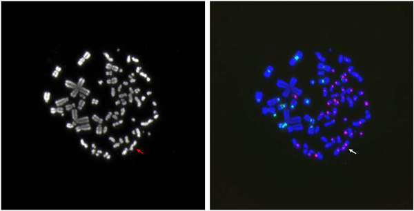
Fig. 10 A dicentric chromosome (arrow) detected by the cell fusion-mediated prematurely condensed dicentric chromosome (PCDC) assay (2-Gy gamma-irradiation).
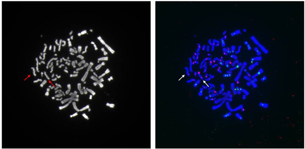
Fig. 11 Dicentric chromosomes (arrows) detected by the cell fusion-mediated PCDC assay(4-Gy gamma-irradiation).
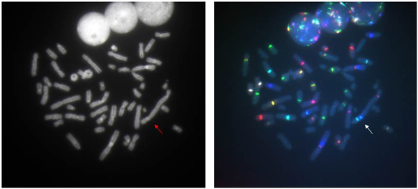
Fig. 12 A tricentric chromosome (arrow) observed in an irradiated human peripheral blood lymphocyte(multi-color centromeric probes, MetaSystems Inc.).
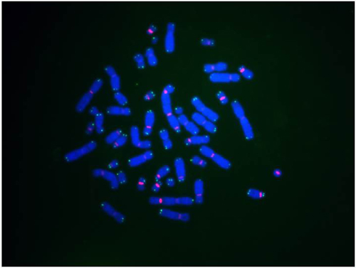
Fig. 13 FISH with centromeric (red) and telomeric (green) PNA probes on a gamma-irradiated human peripheral blood lymphocyte.
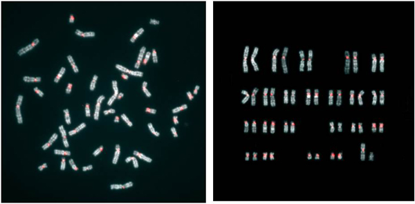
Fig. 14 An example of direct R-banding FISH with the centromeric PNA probe on human peripheral blood lymphocytes.[Cytologia 77: 261-267, 2012.]
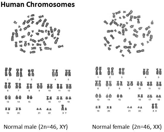
Fig. 15 Human chromosomes.
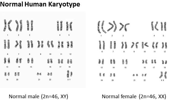
Fig. 16 G-banded chromosomes.
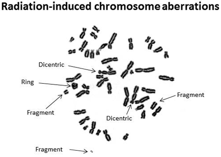
Fig. 17 A human metaphase cell.Peripheral blood lymphocytes were irradiated with 60Co-gamma ray (5 Gy).Dicentric chromosomes, a ring chromosome and fragments were observed.
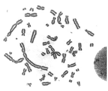
Fig. 18 Chromosome aberrations induced by high-dose (5 Gy) gamma-irradiation.Note that multi-centric chromosomes and fragments were observed.
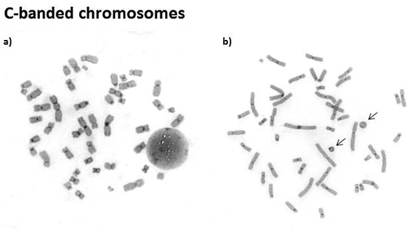
Fig. 19 C-banded chromosomes. All centromeric regions were densely stained with Giemsa by C-banding.
(a)Three dicentrics and three fragments induced by gamma-irradiation.
(b)A centric ring and an acentric ring induced by gamma-irradiation (arrows).
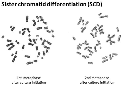
Fig. 20 SCD for analyzing cell kinetics.
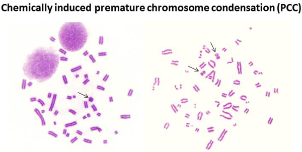
Fig. 21 Examples of PCCs induced by Calyculin-A after 60Co-gamma-irradiation.Arrows indicate ring chromosomes at G2/M phase (5 Gy, left) and G2/A phase (10 Gy, right).
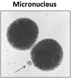
Fig. 22 Micronucleus (arrow).
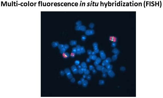
Fig. 23 FISH using a single whole chromosome paint (red) and cloned DNA (yellowish green) probes.Peripheral blood lymphocytes were irradiated with 60Co-gamma ray (5 Gy).Translocations can be detected.
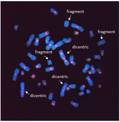
Fig. 24 FISH on gamma-irradiated chromosomes using centromere- and telomere-specific peptide nucleic acid (PNA) probes.Centromeres and telomeres were stained with red and green, respectively. [Cytologia 76: 1-2, 2011, modified]
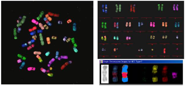
Fig. 25 Multiplex FISH (M-FISH) on human metaphase chromosomes.
Peripheral blood lymphocytes were irradiated with X-ray (2 Gy). 22 autosome pairs and 2 sex chromosomes are painted in different colors and identified.Complex aberrations including translocation can be detected.
Chromosome Aberration Data[PDFファイル/215KB]
Table 1. Dicentric chromosome scoring data of peripheral blood mononuclear cells: Version 1 (30 November 2010).
[Dicentric chromosome assay data collection, version 1]
![]()
PDF形式のファイルをご覧いただく場合には、Adobe社が提供するAdobe Readerが必要です。
Adobe Readerをお持ちでない方は、バナーのリンク先からダウンロードしてください。(無料)
Adobe Reader provided by Adobe is required to view PDF format files.