
Micro-PIXE analyzer
An in-air micro-PIXE (Particle Induced X-ray Emission) analysis system was developed by JAEA (formally JAERI) in 2000. In this system, ion beams are focused down to 1 μm with a precise quadrupole doublet-magnet and scanned over a sample by a couple of electrostatic deflector. Two dimensional distributions of some trace elements in a single biological cell can be measured with spatial resolution of 1 μm.
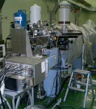
Fig. 1 Micro-PIXE & PIGE analyzer
Principle of PIXE
Hitting by ions with sufficient energy causes inner shell ionization of atoms. When the outer-shell-electrons drop down to the induced inner-shell vacancies, X-rays with energies unique to each element are emitted. By analyzing the energy spectrum of the X-rays, the elements in the specimen can be identified, and also the absolute concentration and distribution of each element can be measured.
 |
1. Hitting an atom using ion beam. |
 |
2. Inner shell ionization. |
 |
3. Dropping down of outer-shell-electrons and Emitting X-rays. |
Fig. 2 Basic principle of PIXE.
Examples of micro-PIXE analysis
Asbestos fiber
Asbestos is called a “silent time bomb” because the asbestos-related symptoms appear after scores of years. Especially, crocidolite and amosite are known to be more lethal. By our micro-PIXE analyzer, the type of asbestos fibers in standard samples can be identified from the spatial distribution of magnesium, as shown in Fig. 3.
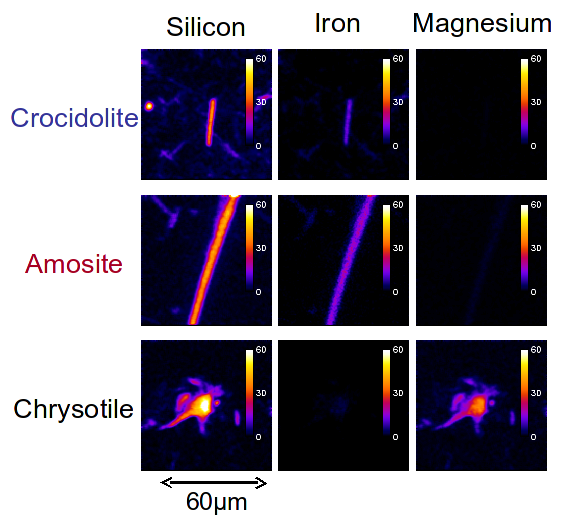
Fig. 3 Analysis results of three kinds of asbestos
High density of magnesium was detected only in the chrysotile.