
100 MeV class microbeam system
A high energy microbeam system with a quadruplet quadrupole magnet lens (Fig. 1) and a set of beam scanner in upstream of the lens has been developed to evaluate response of specific part of intracellular structure such as a cell nucleus, a cytoplasm or cell wall, with a high spatial resolution of 1 μm level.
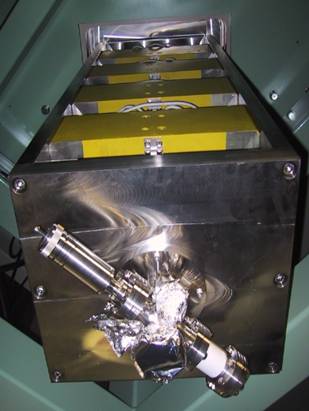
Fig. 1 Quadruplet quadrupole magnet to focus the beams to 1μm level in diameter.
Beam size
A copper-grid (1000 lines per inch) standard sample was used to measure the beam size by secondary electron mapping. Figure 2 shows a typical secondary electron image of the grid obtained with a microbeam of 260 MeV Ne ion. The beam size was estimated for some edges of the grid and mean values were 0.65 μm and 0.67 μm in FWHM for X and Y directions, respectively. These are the finest size of hundred MeV class ion beam in the world.
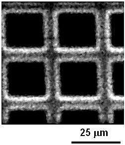
Fig. 2 Typical secondary electron intensity of a copper-grid obtained by 260 MeV Ne microbeam scanning.
Beam fluctuation
Each ion was shot at virtual lattice points on a CR-39 plate with a thickness of 100 μm, as shown in Fig. 3. The beam fluctuations were 0.79 μm and 0.64 μm in FWHM for X and Y directions, respectively.
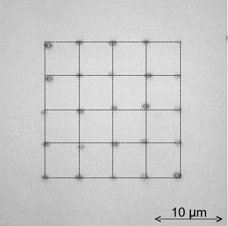
Fig.3 One example of photomicrograph of etched pits in 5 × 5 points made on a CR-39 plate. Intersections of broken lines indicate the target points by beam scanning.