
グループリーダー
小西 輝昭
メンバー
・上原章寛 主幹研究員
・田中泉 主任研究員
・薬丸晴子 研究員
・城鮎美 主任研究員(併任)
・間宮大晴 スチューデントリサーチャー(SR)(立教大学・連携大学院生D2)
・池田真友子 業務補助員
・田中美香 業務補助員
・横地和子 業務補助員
・武田志乃 専門業務員(併任)
受入研究員
・八木唯奈 実習生(大阪大学D2)
・角谷耕太郎 実習生(大阪大学M2)
・霜田宗佑 実習生(大阪大学M1)
・長島湧生 実習生(東京都市大学B4)
研究内容
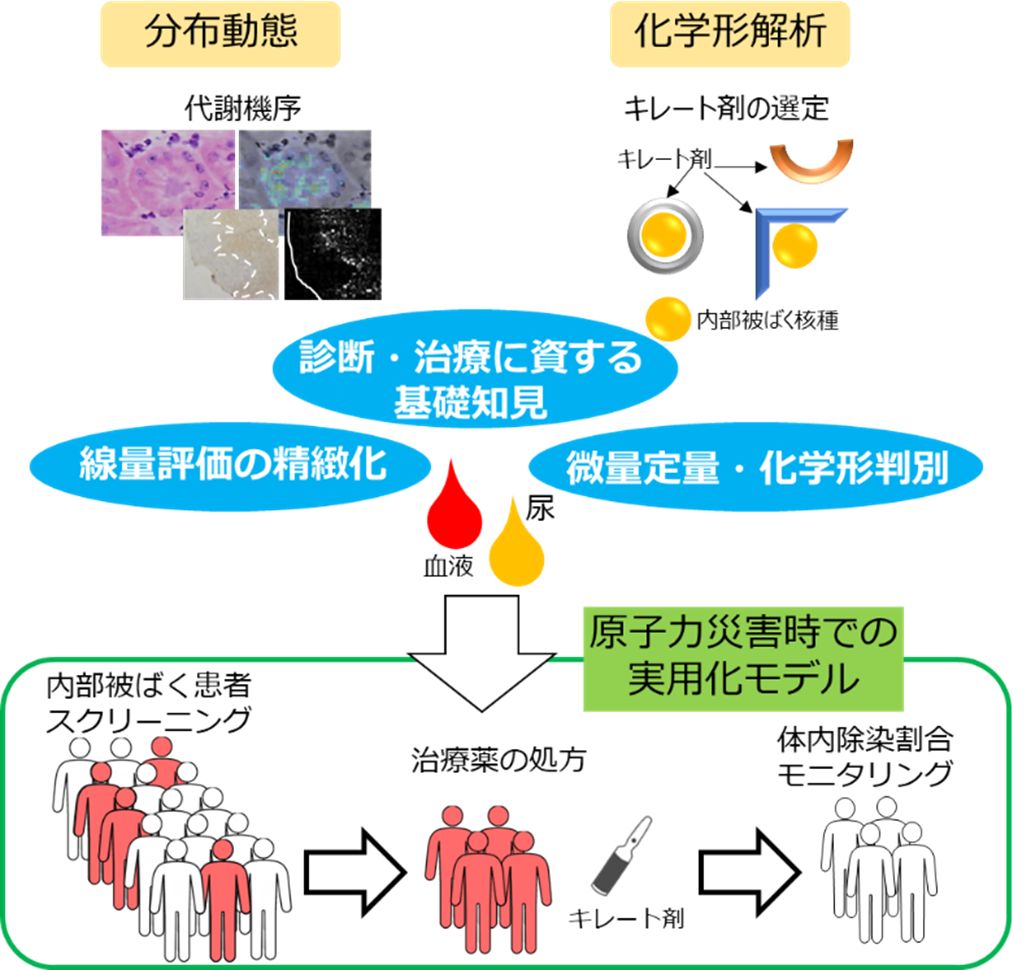
内部被ばく後の除染法や治療技術の高度化研究
私たちはウランなどの放射性核種による内部被ばくの低減化を目指して研究を行っています。
本グループでは、大型放射光施設SPring-8や静電加速器などの先端的な量子ビームを活用したXAFS分析法やマイクロPIXE分析法によって、放射性核種の体内分布、代謝機序および生体内化学形を分子・細胞レベルから個体レベルのスケールで詳細に解析しています。微量元素定量や高精度の被ばく線量の評価が可能になり、適切な治療法の開発につながることが期待されます。
さらに、放射性核種の内部被ばくは、その化学的毒性と放射線障害の両方の作用をもたらすと考えられることから、マイクロビーム細胞照射装置を活用し、様々な放射線種による生物影響についても、細胞レベルでの防御的な細胞応答機構の解明に向けた研究を実施しています。
放射性核種の体外排泄治療薬を用いた除染評価手法を開発し、原子力災害時での内部被ばく患者のスクリーニングおよび体内除染モニタリングに役立てます。この研究成果は、内部被ばくによる体内除染治療に有用な情報を提供します。
陽子線マイクロビーム細胞照射装置(SPICE-QST microbeam)を活用した放射線医科学研究
私たちが研究に活用しているSPICE-QST(Single Particle Irradiation System to Cell)マイクロビームは、3.4 MeVの陽子線を直径約2 µmの極細ビームに集束させ、培養細胞の核や細胞質などの狙った微小部位へ1個単位で正確に照射できる最先端のマイクロビーム細胞照射装置です(図1)。
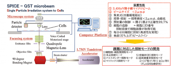
図1 SPICE-QSTマイクロビームの装置概要
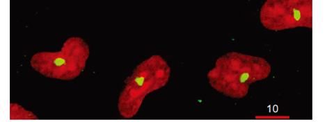
図2 ヒト肺がんA549培養細胞の細胞核(赤)に陽子線500個のマイクロビーム照射を行った。DNA二本鎖切断部位を示すγH2AX蛍光スポットを黄色で示した。
当グループでは、本装置が持つ卓越した単一粒子制御性と位置精度を活かし、個々の細胞を通過する粒子数を厳密に制御する「素線量の放射線生物学」を軸に据えて、従来の統計的予測を超えた低線量域における細胞・遺伝子応答のメカニズム解明に挑んでいます。さらに、装置の開発および高度化にあたっては、量医研・静電加速器運転室との密な連携を図ることで、生物学的な要請に的確に応えるアプリケーションオリエンテッドな高度化を実現しています。
現在、従来の陽子線専用から5.1 MeVアルファ線マイクロビームへと展開するさらなる高度化を進めています。この技術革新により、アルファ線放出核種を用いる標的アイソトープ治療(TAT)や、ウラン・プルトニウムといったアクチニド元素による内部被ばくの影響評価を、シングルセルレベルで解明するための研究開発を視野に入れています。
こうした基盤のもと、量医研との共同研究を通じて、ブラッグピーク近傍の高LETプロトン線が引き起こすがんの免疫逃避メカニズムの解析や、細胞質・ミトコンドリアへの選択的照射によるバイスタンダー効果・適応応答の解明を進めています。また、ライブセルイメージング技術を活用してDNA損傷修復タンパク質の集積と離散動態を単一細胞レベルで定量解析するなど、独自の画像解析アルゴリズムの開発や国際共同研究(IAEA-CRP等)との緊密な連携を交え、単一細胞レベルの微視的物理作用から次世代の放射線治療・防護の高度化へと直結する先駆的な研究を展開しています。
放射線医科学研究基盤高度化のための国際共同プロジェクト
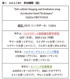
世界15機関による国際連携体制で実施されている国際原子力機関国際共同プロジェクト(IAEA-CRP F11024)に参加しています。
プロジェクト目的は、単一細胞の高分解能イメージングと照準照射を可能にするマイクロビーム照射技術による放射線治療・医科学研究基盤の高度化と研究推進です。
当該グループは、所内外・国内外の研究グループと連携して、陽子線とα線の生物効果を単一細胞レベルで解析し、放射線障害低減および緊急被ばく医療の高度化に資する研究を推進しています。静電加速器棟マイクロビーム細胞照射装置SPICEは、IAEA-CRPにおける放射線生物学研究の重要な役割を担っています。
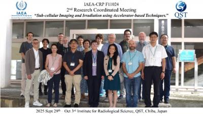
以下は、参加国(機関)リストとQST千葉地区で開催された第二回企画会議の参加者です。
(IAEA-CRP F11024 “Sub-cellular Imaging and Irradiation using Accelerator-based Techniques.” Project Duration 2023 May 19 – 2028 July 1)
↵
List of Papers
- Akiyama D, Uehara A, Kirishima A, Sato N (2026) High temperature reactions of uranium oxide and boron carbide with zirconium oxide under reducing and oxidizing conditions. Journal of Nuclear Materials. 633. DOI: 10.1016/j.jnucmat.2026.157036
- Li C, Liu X, Zhu L, Liu M, Konishi T, Lou X, et al. (2026) Carbon ion irradiation at a clinically used dose rewires super-enhancers to drive necroptosis and systemic immunity. Science Advances. 12(35):eaec7590.
- Kodaira S, Kusumoto T, Mamiya T, Konishi T, Ogawara R, Kamiguchi N, Amano D, Sasai K (2026) Characteristics of passive dosimeters for ultra-high dose rate protons and electrons. Radiat Meas. 2026;192:107629.
- Autsavapromporn N, Mamiya T, Liu C, Duangya A, Tengku Ahmad TA, Oikawa M, Konishi T (2026) Bystander effects in both hypoxic cancer and normal cells under FLASH irradiation using proton microbeams. International Journal of Radiation Biology.102(8): 1001-1013 doi: 10.1080/09553002.2026.2654422:1-13.
- Kusumoto T, Konishi T, Picollo F, Sturari S, Aprà P. (2026) Changes in yields of water radiolysis products in the presence of hydrogenated nanodiamonds under X-rays: Significant increase of hydrogen peroxide. Colloids and Surfaces A: Physicochemical and Engineering Aspects. 737:139871.
- Autsavapromporn N, Mamiya T, Liu C, Duangya A, Tengku Ahmad TA, Oikawa M and Konishi T (2026) Bystander effects in both hypoxic cancer and normal cells under FLASH irradiation using proton microbeams. Int J Radiat Biol. 1-13.DOI: 10.1080/09553002.2026.2654422
- Mamiya T, Kusumoto T, Sasai K, Kamiguchi N, Amano D, Kurita K, Kodaira S and Konishi T (2025) Ultra-high dose rate protons in clinical irradiation suppress DNA single-strand breaks induction in the below nano-second time scale of physicochemical stages. Int J Radiat Biol. 101: 1098-1104. DOI: 10.1080/09553002.2025.2548470
- Kirishima A, Takei M, Uehara A and Akiyama D (2025) Determination of the thermodynamic quantities for complex formation of U(VI) with amino acids in aqueous solution. J. Chem. Thermodyn. 206: 107469.DOI: 10.1016/j.jct.2025.107469
- Uehara A, Tanaka I, Ishihara H, Akiyama D, Kirishima A and Homma-Takeda S (2024) Monitoring method for uranium concentration and chemical form in the droplet of rat serum. J Toxicol Sci. 49: 543-548.DOI: 10.2131/jts.49.543
- Fujii T, Kato C, Wada N, Uehara A, Sossi P and Moynier F (2023) Theoretical and Experimental Study on Vanadium Isotope Fractionation among Species Relevant to Geochemistry. ACS Earth Space Chem. 7: 912-925.DOI: 10.1021/acsearthspacechem.2c00299
- Konishi T, Wang J, Kobayashi A, Liu C, Hiroyama Y, Mamiya T, Kusumoto T and Oikawa M (2025) Microbeam irradiation reveals cytoplasmic damage contributes to micronuclei induction. The European Physical Journal Plus. 140.
- Kusumoto T, Danvin A, Mamiya T, Arnone A, Chefson S, Galindo C, Peaupardin P, Raffy Q, Kamiguchi N, Amano D, Sasai K, Konishi T, Kodaira S (2024) Dose Rate Effects on Hydrated Electrons, Hydrogen Peroxide, and a OH Radical Molecular Probe Under Clinical Energy Protons. Radiat Res. 201(4):287-293.
- Wang J, Oikawa M and Konishi T (2023) Stimulation of Nuclear Factor (Erythroid-Derived 2)-like 2 Signaling by Nucleus Targeted Irradiation with Proton Microbeam. Biology (Basel). 12.
- Konishi T, Kusumoto T, Hiroyama Y, Kobayashi A, Mamiya T and Kodaira S (2023) Induction of DNA strand breaks and oxidative base damages in plasmid DNA by ultra-high dose rate proton irradiation. Int J Radiat Biol. 99: 1405-1412.
- Uehara A, Matsumura D, Tsuji T, Yakumaru H, Tanaka I, Shiro A, Saitoh H, Ishihara H and Homma-Takeda S (2022) Uranium chelating ability of decorporation agents in serum evaluated by X-ray absorption spectroscopy. Anal Methods. 14: 2439-2445. DOI: 10.1039/d2ay00565d
- Kobayashi A, Hiroyama Y, Mamiya T, Oikawa M and Konishi T (2023) The COX-2/PGE2 Response Pathway Upregulates Radioresistance in A549 Human Lung Cancer Cells through Radiation-Induced Bystander Signaling. Biology (Basel). 12.
- Autsavapromporn N, Kobayashi A, Liu C, Jaikang C, Tengku Ahmad TA, Oikawa M and Konishi T (2022) Hypoxia and Proton microbeam: Role of Gap Junction Intercellular Communication in Inducing Bystander Responses on Human Lung Cancer Cells and Normal Cells. Radiat Res. 197: 122-130.
- Ishii S, Toki J, Oikawa M, Ohsawa D (2020) Konishi T. Electrical responses of a carbon nanotube thin-film transistor to MeV proton irradiation in air. Radiation Effects and Defects in Solids. 175(5-6):440-449.
- Dukaew N, Konishi T, Chairatvit K, Autsavapromporn N, Soonthornchareonnon N, Wongnoppavich A (2020) Enhancement of Radiosensitivity by Eurycomalactone in Human NSCLC Cells Through G(2)/M Cell Cycle Arrest and Delayed DNA Double-Strand Break Repair. Oncol Res. 28(2):161-175.
- Wang J and Konishi T (2019) Nuclear factor (erythroid-derived 2)-like 2 antioxidative response mitigates cytoplasmic radiation-induced DNA double-strand breaks. Cancer Sci. 110: 686-696. DOI: 10.1111/cas.13916
- Konishi T, Kodaira S, Itakura Y, Ohsawa D and Homma-Takeda S (2019) Imaging Uranium Distribution on Rat Kidney Sections through Detection of Alpha Tracks Using Cr-39 Plastic Nuclear Track Detector. Radiat Prot Dosim. 183: 242-246.
- Konishi T, Oikawa M, Suya N, Ishikawa T, Maeda T, Kobayashi A, Shiomi N, Kodama K, Hamano T, Homma-Takeda S, Isono M, Hieda K, Uchihori Y and Shirakawa Y (2013) SPICE-NIRS microbeam: a focused vertical system for proton irradiation of a single cell for radiobiological research. J Radiat Res. 54: 736-747.DOI: 10.1093/jrr/rrs132
- 解説記事:小西輝昭. (2025) IAEA国際共同研究プロジェクト:マイクロビーム照射技術の高度化による放射線医科学・がん治療研究への貢献. Isotope News. 797: 18-21.